Deformational Plagiocephaly
Deformational Plagiocephaly
Deformational Plagiocephaly
Deformational Plagiocephaly
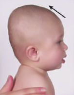
Positional or deformational plagiocephaly is the most frequent type of the cranial shape asymmetry or deformation.
This is the most common form of non-synostotic cranial deformity resulting from persistent unidirectional force exertion over the back of the head. This results in unilateral flattening on the parieto-occipital region which may be accompanied by compensatory volume shift towards ipsilateral frontal bone and on bi-parietal plane which, may in turn lead to ipsilateral prominence of the maxillofacial structures and anterior displacement of the ipsilateral ear.
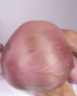
From Above
Deformational Plagiocephaly
Deformational Plagiocephaly
- One sided flattening on the rear region of the skull.
- The shape of the head resembles a parallelogram.
- The ear on the flattened side of the cranium is displaced forwardly.
From Side
Deformational Plagiocephaly
From Side
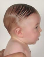
- One of the ear seems to be nearer to the shoulder.
Front
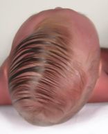
From Below
From Side
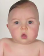
- One eye seems to be smaller related to other.
- One cheek seems to be stuffed when compared to other side.
- One half of the head seems to be taller than the other half.
From Below
From Below
From Below

- Forehead seems inclined not flat.
- Half of the face region seems angulated and inclined.
- Eyes and ears are not in the same plane.
- Half of the face and one cheek seems to be more stuffed as compared to the other side.
Deformational Brachicephaly
Deformational Brachicephaly
Deformational Brachicephaly
Deformational Brachicephaly
Positional or deformational brachicephaly is another frequent type of the cranial shape asymmetry or deformation.
This is also a common form of non-synostotic cranial deformity resulting from persistent force exertion over the back of the head. This results in bilateral flattening on the parieto-occipital region.
When looked from a side, back to front head length seems to be reduced and the forehead seems to be declined backwards.
From Above
Deformational Brachicephaly
Deformational Brachicephaly
- Ear to ear distance is greater than forehead to back distance. Head is wider than normal
- The back of the head is completely flat and lost its curved shape.
From Side
Deformational Brachicephaly
From Side
- Back to front head length seems to be reduced.
- Rear half of the head seems to be taller.
- Forehead seems to be declined backwards.
Front
From Below
From Side

- The face seems to be proportionally smaller as compared to head size.
- The head seems to be wide.
- Head may be widest above the ears.
- The top of the ears seem to be displaced away from the head.
From Below
From Below
From Below

- The middle portion of the head seems to be wider than the face and the chin.
- The widest part of the head is the region above the ears.
Deformational Scaphocephaly

Positional or deformational scaphocephaly is another type of the cranial shape asymmetry or deformation.
This is common especially in premature babies. Premature babies have especially soft skulls and often spend time in the neonatal intensive care unit with the head in a fixed position. Usually the head appears as pressed from both sideways and seems longer, taller and narrower than normal.
When looked from the front, head length between the ears seems to be reduced and the forehead seems to be narrow.
PREVENTING AND TREATMENT OF CRANIAL SHAPE DEFORMITIES

In mild cases or in very early stages of deformation some simple preventive measures may be applied under supervision of a medical specialist. These include:
- Active head repositioning during sleep time.
- Changing the sleeping position.
- Encouragement of ‘tummy time’ as to decrease time spend in back laying position when awake.
In moderate and prominent cases or in patients refractory to those conservative measures helmet therapy can be instituted.
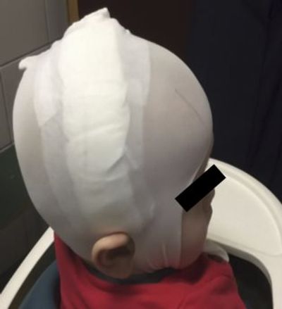
Helmet Treatment

In cases of deformational cranial shape deformities helmet therapy can be instituted under the supervision of a specialist. Therapy depends on the cause, the age of the child and the severity of the head deformity. Helmet application was first described in 1979. In recent years, technological proceedings yielded precise 3D measurements and finest modelling and the success rates of helmet therapy increased significantly.
Nowadays, molding helmet therapy using an individual (custom made) cranium orthosis presents a widely accepted treatment option for infants with positional head deformities.
OUR POLICY IN HELMET TREATMENT
Diagnosis and indication should be supervised by a specialist (preferably a pediatric neurosurgeon). In our practice, we perform our work according to the expertise, opinions and suggestions of the medical specialists in each step. Testing the suitability of the produced helmet and follow up evaluations are all performed under the supervision of a pediatric neurosurgeon.
PRINCIPLES OF CUSTOM MADE HELMET THERAPY
The rationale behind helmet therapy is structured on the fact that skull enlargement will proceed towards the area with the least resistance. Cranial orthoses can be classified as active or passive helmets depending on any pressure applied to protruded parts of the skull. Active cranial orthoses work via applying firm pressure over the bulged parts of the skull whereas in passive helmets a potential space is left behind the effected region with other parts staying in close proximity to the skull without applying pressure. In other words, passive orthoses allow room for growth in the flattened areas while minimal pressure is applied to the areas with bossing, whereas active orthoses apply compression to the bossed areas, possibly resulting in a more rapid deformity correction.
SUCCESS OF HELMET THERAPY

In clinical studies based on positional cranial deformation cases, a correction rate of 81% was achieved using helmet therapy irrespective of deformity severity [1,3,5]. Improvement of asymmetry was found to be significantly better in patients in whom, helmet therapy has been started earlier than 6 months of age [2,4,6].
In our photography gallery, you can see the pictures of our cranial shape deformation cases which have been treated with professional custom-made CH-Pro (A) active helmet system.
CRANIOSYNOSTOSIS
A baby’s skull is made up of several large bones. The places where these bones touch are flexible connections called sutures. The location where four of these large bones meet in the front of the head is called the anterior fontanelle, or soft spot (there is another one in the back of the head). This flexibility allows the head to fit through the birth canal, and permits the brain to grow. The brain quadruples in size the first two years of life, and the bones of the skull must grow and not restrict its growth. Bone growth occurs at these sutures.
When a suture is not formed or closes too soon, it is called craniosynostosis. It is estimated that this defect occurs in one out of every 2500 live births. Craniosynostosis causes the head shape to be deformed, and in certain instances, can prevent the brain from having enough room to grow.
The specific abnormality of the head shape depends on which suture(s) is closed or fused. An abnormal head shape is noticed after birth. One needs to distinguish whether an abnormal head shape in an infant is primarily due to craniosynostosis or associated with positional molding in terms of the appropriate treatment and in order to avoid the sequels that can be encountered in the long term.
Treatment of craniosynostosis is surgical. Treatment for craniosynostosis is required to prevent the psychosocial implications of having a major deformity and in many cases to prevent elevated brain pressure. Type of the surgical procedure differs due to severity of the cranial shape deformation and to the number of the sutures affected. The surgery should be done by a pediatric neurosurgery team.
STEPS IN CRANIOSYNOSTOSIS TREATMENT

Different surgical techniques are used in craniosynostosis treatment. Sort of the surgical technique is decided according to the type of the craniosynostosis and to the age of the patient.
Surgical reconstruction of the cranial bones and sutures are performed to achieve normal development of the skull and the brain.
In recent years, endoscopic strip craniectomy or suturectomy has gained acceptance as a minimally invasive technique in craniosynostosis surgery with complementary helmet therapy. Endoscopic procedures are preferably performed for simple non-syndromic craniosynostosis cases with single suture synostosis and postoperative use of custom made helmets in those cases usually yields to satisfactory results. Besides, recent reports have shown that cranial-orthotics may also play an important role in post-surgical molding and maintenance of the cranial shape following cranial vault remodeling in both simple and complex craniosynostosis cases which have been operated with classic techniques.
BASIC FEATURES OF THE HELMET TREATMENT
The rationale behind helmet therapy is structured on the fact that skull enlargement will proceed towards the area with the least resistance. Cranial orthoses can be classified as active or passive helmets depending on any pressure applied to protruded parts of the skull.
Active cranial orthoses work via applying firm pressure over the bulged parts of the skull whereas in passive helmets a potential space is left behind the effected region with other parts staying in close proximity to the skull without applying pressure.
In other words, passive orthoses allow room for growth in the flattened areas while minimal pressure is applied to the areas with bossing, whereas active orthoses apply compression to the bossed areas, possibly resulting in a more rapid deformity correction.
In craniosynostosis cases, usually passive helmets are recommended. Following craniosynostosis surgery, passive helmets help to protect the surgically reconstructed skull and act as a guide to keep the cranial growth normally as well.
HELMET TREATMENT FOLLOWING CRANIOSYNOSTOSIS SURGERY

Following craniosynostosis surgery, custom made helmets can be used as a support to surgically reconstructed skull and as an orthotic guide to keep the cranial growth normally.
In recent years, it has been shown that custom made helmet therapy is significantly effective for achieving satisfactory cosmetic results when used as an adjunctive treatment following craniosynostosis surgery in children. Regarding this issue, a clinical study had been published in 2015 and the advantages of custom made helmet use were underlined in detail [8].
At the international congress of ISPN (International Society of Pediatic Neurosurgery) which was held at Japan in year 2016, a scientific study presented the results of custom made helmet use following the surgical treatment of craniosynostosis cases. The results of the study showed that helmet use increased the effectivity of surgical correction significantly [7].
CRANIO-HEALTH “CP-PRO HELMET” PRODUCING STANDARDS IN CRANIOSYNOSTOSIS CASES
Since we give our best effort to reach the finest quality, we always keep eye on the latest technological and scientific proceedings. Our main target is to give the best and the healthiest assistance to our patients during the infancy period when the development of the skull and the brain is most sensitive, and hence, requires the most careful care and utmost responsibility.
The production of custom-made helmets first requires a precise measurement process by reliable software. The second step is the moulding of the helmet material to the exact volume and shape that will suit the patient’s skull growth potential which, requires a meticulous production process.
Our helmet producing procedure involves the supervision of a “pediatric neurosurgery specialist” as a gold standard. Both the measurement and the first application steps are done under control of the medical specialist. Regular clinical follow-up control examinations are performed by the medical specialist and the period of helmet application therapy is decided by the pediatric neurosurgeon, preferably by whom the indication has been given.
In order to provide the most accurate and healthy assistance to our patients, we meticulously perform our work according to the expertise, opinions and suggestions of medical specialists in each step.